Services
Ultrasound
Diagnostic ultrasound, also called sonography, is a simple, safe and painless imaging method that uses high-frequency sound waves to produce images of the structures inside your body
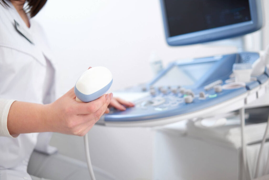
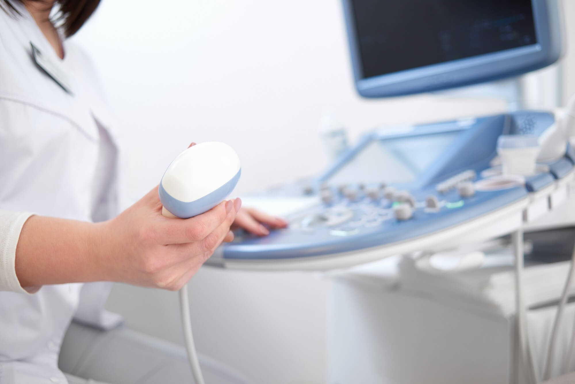
Our radiologist or sonographer will apply a conducting gel to your skin and use a transducer to pass the sound waves through your body. The ultrasound unit captures these “echoes” to create an image that is instantly displayed on a monitor. These images can then be printed.
Ultrasound is useful for the evaluation of solid organs in the abdomen (e.g., the liver, spleen, and kidneys), for breast abnormalities, and for examining arteries and veins using the Doppler ultrasound technique to assess flow and obstruction.
There is no injection required or radiation exposure associated with ultrasound procedures. The gel may feel cold on your skin.
Ultrasound is used as a supplementary tool for guidance with aspirations and biopsies.
Certain ultrasound examinations require specific preparation.
- A full bladder is required for pelvic ultrasound. Sound waves travel more easily through liquids than through surrounding tissues and air. Consequently, a full bladder facilitates the passage of sound waves through it, making the uterus and ovaries behind the bladder easier to see.
- Avoid eating or drinking anything 4 – 6 hours before your abdominal ultrasound. When you eat, drink, or even chew gum, you introduce air into the stomach and bowel. Sound waves do not penetrate air, making it harder to see the target organs. Food can also make your gallbladder contract, making it harder to image.