Services
MRI
Magnetic Resonance Imaging (MRI) is a non-invasive test that produces detailed, highly diagnostic imaging of the internal organs. Instead of ionising radiation, it uses a very powerful magnetic field and radio frequency pulse waves to record the rotating magnetic field produced by atoms in the body.




Magnetic Resonance Imaging (MRI) is a non-invasive test that produces detailed, highly diagnostic imaging of the internal organs. Instead of ionising radiation, it uses a very powerful magnetic field and radio frequency pulse waves to record the rotating magnetic field produced by atoms in the body.
gown. You will also need to remove all jewellery (including wristwatches and body piercings) and any other metallic objects. It is preferable that you leave items of value at home. There is no need to stop medication for the examination. You may eat and drink normally unless instructed otherwise.
You need to complete a consent form. Your healthcare provider or radiographer will explain the procedure and its risks.
Before your MRI scan, tell your doctor and the MRI radiographer if you:
- Are allergic to any medicines. The contrast material used for MRI does not contain iodine. If you have a known allergy to the contrast material used for MRI, tell your doctor before having the test. Sometimes, the benefits of having this test may outweigh the risks.
- Are pregnant or breastfeeding? The contrast agent is excreted into the breast milk, so breastfeeding should be discontinued for 24 hours.
- Have a pacemaker, artificial limb, any metal pins or metal parts in your body (especially in the eyes), metal heart valves, metal clips in your brain, metal implants in your ear, tattooed eyeliner, or any other implanted or prosthetic medical device (such as a medicine infusion pump).
- Have had an accident or work with metal. This increases the possibility that you have metal fragments in your head, eyes, skin, or spine. An X-ray may need to be taken first to determine whether the MRI scan is safe.
- Had recent surgery (last 3 months) on a blood vessel. In some cases, you may not be able to have the MRI test.
- Are claustrophobic. You are required to lie still inside the MRI and may need medicine to help you relax.
- Have any other health conditions, such as kidney disease that may prevent you from having an MRI using contrast material. Contrast agents for MRI contain Gadolinium and are an extremely safe group of drugs. Side effects rarely develop. Relative contraindications would be allergy to Gadolinium or poor kidney function. Therefore, depending on age and risk factors, you may be asked to do a blood test to assess kidney function prior to the MRI scan.
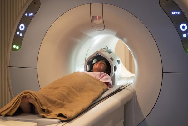
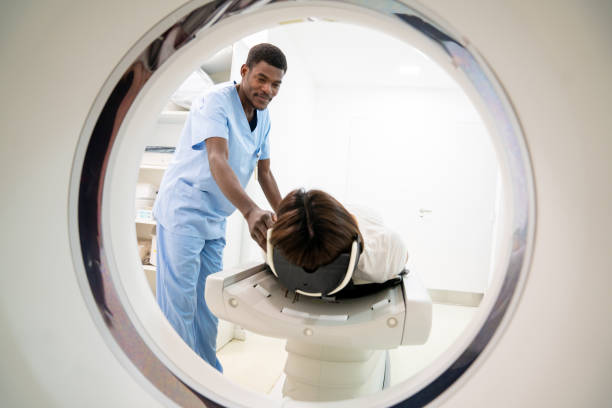
You will be asked to lie on an examination bed, which will be raised up and into the magnetic field. You will find yourself in an open-ended cylinder just over 1 meter in length. It remains open at both ends for the entire duration of your scan. The cylinder is well-lit and ventilated by a fan.
Once in position, it is most important that you remain as still as possible as movement detracts from the quality of images obtained. You will hear a variety of loud noises, but we will provide you with either earphones or earplugs (depending on the type of examination) to compensate for this. Most examinations typically run for 30 minutes, although some scans may last for up to an hour.
The radiographer will be in contact with you at regular intervals during the scan to ensure that you are still comfortable and to let you know how long each part of the scan will take.
For some examinations, a contrast agent is required to provide a complete diagnosis. This is a substance called Gadolinium, and it is injected into a vein. It is safe and painless. The radiographer will, wherever possible, talk to you about this. Apart from intravenous contrast, certain studies also require oral contrast. The procedure for oral contrast will be explained prior to the study. There are also particular examinations that require a period of fasting for approximately 6 hours prior to the test.
Some specific examinations do require what we call breath-hold intervals. The radiographer will explain and guide you thoroughly through this.
Occasionally, an MRI arthrogram may be necessary to visualise the joint structures. These cases require the injection of contrast material and sterile saline directly into the joint before the MRI. This can be done using fluoroscopic or ultrasound guidance.