Services
Fluoroscopy
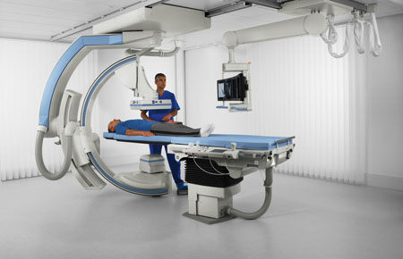
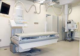
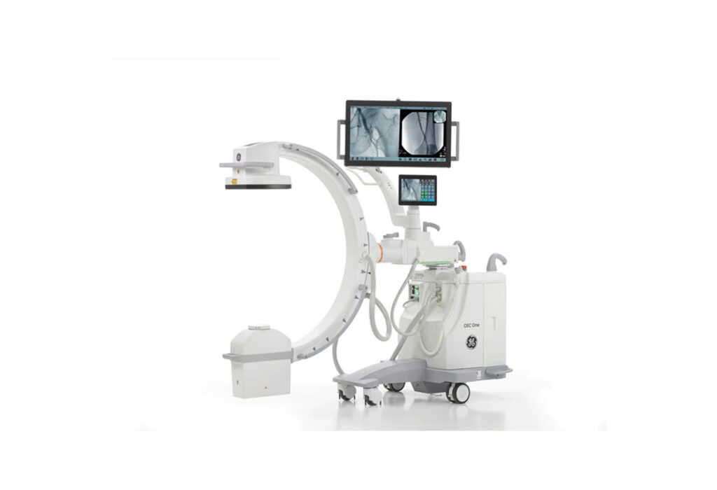
Fluoroscopy is a type of medical imaging that shows a continuous X-ray image on a monitor, much like an X-ray movie. During a fluoroscopy procedure, an X-ray beam is passed through the body. The image is transmitted to a monitor so the movement of a body part or of an instrument or contrast agent ("X-ray dye") through the body can be seen in detail.




Fluoroscopy is a radiologic modality that provides real-time X-ray imaging, especially useful for guiding a variety of diagnostic and interventional procedures.
Fluoroscopy may be part of an examination or procedure that is done on either an outpatient or inpatient basis. The specific type of procedure or examination being done will determine whether any preparation prior to the procedure is required. Your doctor should notify you of any pre-procedure instructions.
A barium enema is a fluoroscopic examination of the large intestine where the entire cavity of the colon is filled with barium, and abdominal X-rays are taken.
How the Test is Performed
This test is done after the colon is completely empty. Your doctor will give you instructions on how to empty the colon.
You will lie on the X-ray table, and an X-ray will be taken.
You will then be told to lie on your side. The healthcare provider will gently insert a well-lubricated tube (enema tube) into your rectum. The tube is connected to a bag that holds a liquid containing barium sulphate. A small balloon at the tip of the enema tube may be inflated to help keep the barium inside your colon. The healthcare provider will monitor the flow of the barium on an X-ray fluoroscopy screen.
You will be asked to move into different positions, and the table will be slightly tipped to get different views. At certain times when the X-ray pictures are taken, you will hold your breath and be still for a few seconds so the images are not blurred.
The enema tube will be removed after the pictures are taken. You will be given a bedpan or helped to the toilet, so you can empty your bowels and remove as much of the barium as possible. One or two X-rays may be taken after you use the bathroom.
arium Swallow, Meal, and Small Bowel Follow Through
An upper GI and small bowel series is a set of X-rays taken to examine the oesophagus, stomach, and small intestine.
How the Test is Performed
You may be given an injection of a medication that will temporarily slow muscle movement in the small intestine so structures can be more easily seen on the X-rays.
Before the X-rays are taken, you must drink a cup of a milkshake-like drink that contains a substance called barium, which shows up well on X-rays.
Fluoroscopy tracks how the barium moves through your oesophagus, stomach, and small intestine. Pictures are taken with you in a variety of positions. You may be sitting or standing.
The test can take 30 minutes for a Barium swallow or can be up to 3-6 hours for a complete follow-through of the intestines.
How to Prepare for the Test
You may be told to change your diet for the test.
Be sure to ask your healthcare provider if there are any medication restrictions. Generally, you can continue taking medications you take by mouth. Never make any changes in your medications without first talking to your healthcare provider.
You will be asked to remove all jewellery on your neck, chest, or abdomen before the test.
How the Test Will Feel
The barium drink may cause mild bloating but usually causes no discomfort.
A Voiding Cystourethrogram (VCUG) is an X-ray examination of a patient’s bladder and lower urinary tract that uses fluoroscopy and a contrast agent. A VCUG enables the radiologist to detect abnormalities in the flow of urine from the bladder through the urethra. The examination is requested in paediatric and adult patients.
How the Test is Performed
The radiographer begins by positioning you on the table. Infants and young children may be wrapped tightly in a blanket to help them lie still during the imaging. An X-ray of the abdomen is performed.
After cleaning the genital area, a catheter is inserted through the urethra (the tube that carries urine from the bladder out of the body) into the bladder. Then, the bladder is filled with a water-soluble contrast material. Using fluoroscopy, the radiologist will obtain images of your bladder filling and emptying. The radiologist and/or the radiographer will check to see if any of the liquid goes backward into one or both ureters and whether the shape and contour of the bladder and urethra are normal.
When the bladder is full, the radiologist will remove the catheter. We will ask you to urinate into a container. This may be difficult, especially for children but it’s important. Babies will urinate on their own when the bladder is full. X-rays will be obtained during the monitoring.
When the examination is complete, you will be asked to wait until the radiologist determines that all the necessary images have been obtained. A voiding cystourethrogram is usually completed within 30 minutes.